


Chest osteochondrosis is a common degenerative disease. There are specific symptoms of thoracic osteochondrosis, which indicate the onset of pathology. In the early stages, the discomfort does not bother the patient much, so he is in no hurry to seek help from a specialist. Over time, the symptoms worsen, forcing the patient to go to the doctor, where there is a neglected pathology. You should know what early signs of osteochondrosis are determined, and what treatment methods are most effective.
What is thoracic osteochondrosis and how does it arise
Osteochondrosis of the thoracic region is characterized by the occurrence of destructive-dystrophic processes in the middle part of the ridge. The destruction is located between vertebrae 8 and 19To find out which vertebra is affected, an accurate diagnostic study needs to be done. Osteochondrosis of the thoracic region is often accompanied by very severe complications, including prolapse or hernia. Without complications, the disease is rare, as the destruction of cartilage tissue will inevitably lead to the destruction of the entire vertebral skeleton.
When a patient experiences circulatory disorders or age -related joint wear, the fibrous ring located in the intervertebral disc cavity begins to collapse, losing its normal structure. Because its destruction is slow, then in the early stages microcracks appear, in which the nucleus pulposus permeates.
As the internal components secrete, the annular fibrosus begins to weaken, causing it to stretch and rupture gradually. When the nucleus pulposus protrudes, an intervertebral hernia occurs, which is the most common complication of osteochondrosis. Pathology involves damage to cartilage tissue, which causes significant discomfort. Severe back pain is also associated with neurological syndromes that arise as a result of pinching or irritation of nerve roots.
Symptoms of breast osteochondrosis
In the early stages, the patient does not feel uncomfortable, therefore, at this stage, the disease can only be detected by chance. The disease has many symptoms that can be disguised as other pathologies.
Symptoms of chest osteochondrosis can be felt by the following manifestations:
- Breathing hard. Problems arise, indicated by shortness of breath and a feeling of shortness of breath. This indicates damage to the spine and spinal cord.
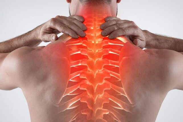
- The main symptom is pain in the chest area. There is also a depressing feeling in the liver, which is reminiscent of an ischemic attack.
- Discomfort occurs when the back is bent. As the disease progresses, the pain in this position increases.
- Against the background of deteriorating blood circulation, there is a feeling of coldness in the lower or upper legs.
- Pain in the chest with a background of appearing intervertebral hernia. Discomfort is often felt on the left or right side of the affected area.
- Throat discomfort and swallowing problems. If there is irritation of the nerve endings in the upper part of the thoracic region, a cough appears.
- Women may experience chest pain that is not related to cyclical changes or hormonal imbalances.
- A tingling or burning sensation appears in the feet and legs.
- Hair and nails become brittle, dull.
- Herpes zoster is rare.
- Pain in the back and chest occurs at the same time.
- Less commonly, there is discomfort in the stomach, liver, or pancreas.
- The onset of stiff pain in the ribs, indicating intercostal neuralgia.
- There are signs of chest chondrosis and compression - similar pathologies.
- There are problems in the work of the gastrointestinal tract. Nausea, heaviness in the stomach.
- In men, a slight libido may decrease. Problems arise in the genitourinary field.
- When standing or sitting for long periods of time, severe discomfort occurs.
- There is a severe headache accompanied by dizziness. Migraines with aura may appear.
- Patients often experience intercostal neuralgia.
- The pain can radiate to the neck or lower back.
If there is in the thoracic osteochondrosis aggregates and signs or some of them, it is necessary to immediately consult a therapist, neurologist, orthopedist. Also, such symptoms should be observed if there are no problems with the gastrointestinal tract, cardiovascular system and lungs.
There are also acute and subacute symptoms. If, with exacerbation of osteochondrosis of the thoracic region, the patient experiences severe pain so that the patient is unable to work, and he can only see rest in bed, then the subacute course is slow and does not significantly limit the patient's motor activity.
A clear sign of a damp lesion - no acute pain. Symptoms in the subacute stage are removed. There is no discomfort with basic body movements, including inhaling, sneezing or turning. A person does not experience pain in dreams, so the process of falling asleep is facilitated.
In order for subacute disease not to worsen and disappear, important rules must be followed:
- No weight lifting.
- You can't bend sharply.
- It is forbidden to be in a sitting or standing position for a long time. Someone unconsciously in this condition considers the posture dangerous for the spine, so there is an excessive load on the ridge, which causes other deterioration.
- Avoid hypothermia. It has been proven that non -compliance with a comfortable temperature regime for the body turns into an exacerbation of the inflammatory process. Moisture is also harmful to the joints.
The duration of the subacute course is individual. If you follow the medical recommendations, the patient will completely eliminate the discomfort in 2-3 weeks. If conservative treatment and rest do not help, and the patient begins to experience nausea, dizziness and weakness, then it is necessary to immediately see a specialist. Such symptoms indicate recurrence.
The stage of development of osteochondrosis of the thoracic region
There are 4 clinical stages of the disease, during the onset of the patient experiences pathological signs:
- In the early stages, there are no clinical symptoms. The first stage occurs against the background of the appearance of destructive processes on cartilage and bone tissue. In the first stage, there is also no rupture or stretching of the fibrous ring, so there is also no hernia. They can detect early protrusions and signs of cartilage degeneration.
- The second stage indicates minor pain or discomfort. A concerned patient seeks a doctor, therefore, osteochondrosis of the thoracic region is immediately detected. People who do not want to visit a specialist can still undergo a second stage, using available medications, but self -treatment will not be enough for a long time. At this stage, the most common neurological symptoms may appear, including headache, burning in the interscapular zone, pain in the neck, and blood pressure surges. Also at this stage, there is an increase in degenerative destruction in the spine: a protruding fibrous ring, which leads to the appearance of an intervertebral hernia in the thoracic region.
- The third stage is already difficult for the patient. Neurological syndromes continue to develop, including persistent pain in the shoulder blades, arms, collarbone and lower back. Patients may show somatic and autonomic disorders, which indicate disturbances in the function of the nervous system. Patients are often tormented by nausea, relentless headache, dizziness, back pain. Hidden signs of heart, gastroenterological, or lung disease may also appear. At this stage, there is active demineralization of bone and cartilage tissue. There is a tendency for injury.
- The last rank is fourth. Against the background of osteochondrosis and hernias, irreversible consequences arise - the mobility of the intervertebral disc is completely lost, and cartilage tissue at the site of prolonged inflammation is replaced by osteophytes. To remove it, an operation is required.
In order not to run the body into a state similar to level 3 or 4, it is better to see a doctor at all. The sooner the disease is detected and therapy is started, the sooner the patient will return to normal and learn to live with osteochondrosis. The process of pathological destruction cannot be completely stopped, but can be slowed down by leading a healthy lifestyle, using medications and doing daily exercise. The more patients turn to a doctor, the harder it is to stop the severe pain syndrome associated with cartilage tissue degeneration.
Risk factors and causes of disease
There is no exact cause of destructive changes in the spine. An important role in the appearance of pathology is associated with hereditary factors. It has been shown that individuals who experience physical inactivity are more likely to have problems with ridges than those who exercise regularly. Also, excessive physical activity can provoke cartilage destruction at an early age.
Thinning and destruction of the intervertebral disc is closely related to spinal excess. If the muscles are not strong enough, and the buttocks are overloaded, cartilage tissue damage occurs.
What causes osteochondrosis:
- Obesity. When you are overweight, there is a strong heavy pressure on the spine. As a result, premature destruction of bone tissue occurs.
- The presence of anomalies in the structure of bone and cartilage. Such problems are housed even in the period of intrauterine development.
- Congenital asymmetry of the intra-articular gap in the intervertebral joint of the type of tropism anomaly, contributes to the occurrence of degenerative-dystrophic processes in the spine.
- The presence of muscle spasms, spondylosis, chronic persistent trigger points and vascular disorders in the thoracic region. This pathology also contributes to the emergence of osteochondrosis of the thoracic region.
- Prolonged exposure to vibration in the spine in a sitting position. An example of a job is a bus or bus driver.
- Frequent physical strain associated with weight lifting. Examples are working as a loader or a professional sports activity.
- Smoking and alcohol abuse. People with unhealthy lifestyles tend to have mineral deficiencies in their bodies and poor circulation, which causes back problems.
- Inactive lifestyle. With insufficient physical activity, accelerated calcium depletion occurs, which is associated with poor metabolic processes. As a result, the bones become brittle. Also, atrophy of muscle tissue, because the load on the spine increases a lot. The result is pain, frequent discomfort with minimal physical activity.
Due to the intervertebral disc, adequate ridge mobility is provided. The intervertebral disc plays a role as a shock absorber. With the development of osteochondrosis, an accelerated demineralization process takes place, essential moisture from the joints is lost. This causes discomfort, decreased mobility in the spine.
Risk factors for breast osteochondrosis include:
- Old age. In the elderly, natural degeneration occurs, therefore, after 40 years, the disease is detected more often.
- Women. In girls, there are periods that contribute to the active release of calcium from the bones - pregnancy and menopause. Without adequate pharmacological support, spinal disease tends to occur.
- The presence of hormonal disorders, endocrinological diseases. If a patient has diabetes mellitus or hypothyroidism without compensatory, intervertebral disc degeneration may occur at an early age.
- Prolonged immobilization. If the patient is ill and has to lie down for a long time, an atrophic process occurs in the muscles, which causes back pain.
- Previous back injury. When ligaments and tendons are stretched, the risk of osteochondrosis in the thoracic region increases.
- The presence of scoliosis. Poor posture in the future causes serious spinal problems, including osteochondrosis and hernias.
Diagnosis of thoracic osteochondrosis
If the patient suspects a back problem, it is necessary to see a therapist. The doctor conducts a general examination of the patient, asks about complaints, measures blood pressure. If there is a suspicion of a neurological problem, the patient is referred to a narrow specialist - a traumatologist, neurologist or orthopedist.
At an appointment with a specialist, they also ask about complaints, make an initial diagnosis of the patient. Based on the visual inspection, a set of diagnostic measures is set, including:
- Radiography. With the help of X-rays, you can assess the state of the skeletal system in general. If the patient has a hernia or osteochondrosis, pathological indications can be observed - the distance between the intervertebral discs will be reduced, and darkness is sometimes observed at the location of the suspected hernia. If the picture results do not suit the specialist, you should continue to look for the cause of pain and discomfort.
- CT or MRI. The most accurate diagnostic method that allows you to accurately examine the state of the inflammatory focus in the picture. A more detailed picture can be seen on an MRI, but if there are contraindications (presence of a pacemaker or prosthesis in the joint), computed tomography is prescribed. CT is an improved X-ray that allows you to see in detail the bones, tendons and ligaments. The image renders the image in the form of a three -dimensional image, so that the details of the damage can be clearly seen.
- Biochemical and general blood tests. This analysis is needed to assess the health of the patient. If an increase in leukocytes, ESR is found, then this indicates an active inflammatory process in the body. With active destruction of bone tissue, reduced calcium levels and a deficiency of cholesalciferol (vitamin D3) are found in the blood.
- Spinal scintigraphy. Research methods show active destruction of bone tissue. Weak bone tissue is very susceptible to fragility. This method will show the tendency and signs of degeneration.
To diagnose the disease, you need to consult an experienced specialist. For a final diagnosis, a complete clinical picture is required, taking into account several laboratory research methods.
Thoracic osteochondrosis of the spinal space requires differentiation along with the following pathologies:
- Dishormonal spondylopathy.
- Pathology of the urinary system, including urolithiasis, cystitis or pyelonephritis.
- Diseases of the cardiovascular system, excluding sinus arrhythmias, tachycardia and angina pectoris.
- Diseases of the gastrointestinal tract, including chronic pancreatitis, stomach and duodenal ulcers, irritable bowel syndrome.
- Previous injuries, fractures.
- Tumors in the chest, including malignant diseases.
- Rheumatoid arthritis (determined by blood tests for C-reactive protein, rheumatic tests and ESR).
- Osteomyelitis of the spine.
- Acute inflammatory process.
- Ankylosing spondylitis.
- Spondylolisthesis.
Treatment of osteochondrosis of the thoracic spine
To slow the progression of the disease, an integrated therapeutic approach is needed. In the early stages, only conservative therapy is indicated, which consists of the use of medications and physiotherapy treatment methods. In further cases, when the patient has a large hernia and an obvious degree of bone degeneration, surgery is prescribed. Do not self -medicate at home. Folk remedies do not eliminate osteochondrosis of the thoracic spine.
In what cases is surgery performed?
Launching osteochondrosis of the thoracic region has a negative impact on the quality of life of patients. If the patient experiences persistent discomfort that interferes with normal life, considering the lack of effect of drug treatment, then a surgical solution to the problem can be offered.
Absolute indications for surgery include:
- Lack of sensitivity in the bladder and intestines.
- If the sensitivity in the foot is lost and the patient loses the ability to move freely.
- Paralysis due to strong hernia growth.
In other cases, the patient decides to remove the hernial formation independently. If the disease really brings severe suffering and the patient's condition does not improve against the background of conservative treatment, the doctor recommends surgery.
Drug treatment of osteochondrosis of the thoracic spine
During the period of exacerbation, the attending physician prescribes various medications that are needed to be used to relieve the inflammatory process. The acute period is characterized by severe pain that can only be overcome with medication. If the medication intake is adequate, the patient will get better. Only an experienced specialist can prescribe medication; self-medication is not acceptable.
Osteochondrosis of the thoracic spine is treated with the following medications:
- Non-steroidal anti-inflammatory drugs, painkillers, or analgesics. This medication is designed to quickly relieve back pain associated with active inflammatory processes. The effects of taking pills or injections are felt the next day. Taking drugs from the NSAID group is accompanied by side effects with prolonged use, therefore, experts recommend limiting the use of drugs to a minimum period, no more than 1-2 weeks. The most dangerous painkillers for the gastric mucosa, causing gastropathy and inflammation. Patients at risk are given certain medications designed to protect the gastrointestinal mucosa. Examples are proton pump inhibitors, histamine H2 receptor blockers, antacids. People with ulcers and gastritis are better off avoiding the use of NSAIDs or taking modern analogues with selective effects.
- Relax the muscles. This medicine is very effective in treating muscle spasms. Relieves pain associated with muscle tension. They act on trigger points located in the pinched muscle tissue. The more people train, the higher their number. Muscle relaxation can relieve the feeling of tightness in the muscles, and thus show an analgesic effect. You need to take the drug in the course, the average duration of therapy is at least 2-4 weeks.
- Vitamins of group B. Prescribe B1, B6, B12 in the form of injections with a combined composition. In large doses, this substance has an analgesic effect and has a positive effect on the nervous system. Neurotropic drugs are effective for treating pain associated with pinched nerve roots. With the help of nutrition, it is impossible to replenish the norms of these substances necessary to achieve a therapeutic effect, therefore they are prescribed in the form of medicines. The average length of one injection is 2-3 weeks. Then, if necessary, they switch to tablets.
- Anti-inflammatory ointment, gel. If pain is tolerable, and a systemic form of NSAID is contraindicated, external medication is prescribed. The advantage of an external remedy is that it does not cause side effects. In rare cases, skin allergies may appear, but the ointment will not cause gastrointestinal or laboratory bleeding. Another advantage of external products is the possibility of long -term use. You can rub the gel for up to 4 weeks, after which rest for a while. The scheme and duration of therapy are determined by the attending physician.
- Honroprotectors. This is a complex material used to nourish the cartilage tissue in the joints. Need to use the drug for a long time, at least six months, after which they rest 2-3 months and the course of therapy is repeated. In 2-3 months, the injectable release form is used, as it is better absorbed. Then they switched to supportive treatments, including the use of tablets. It is important to understand that drugs do not stop the destruction of cartilage tissue. They only produce extra nutrients, which slow down the degenerative processes that take place in the bones and joints.
- Inventory of calcium and vitamin D3 complexes. It has been proven that residents in the northern latitudes do not get enough vitamin D3, due to low solar activity throughout the year in the region. To get rid of hypovitaminosis, it is necessary to take cholecalciferol supplements in winter and autumn in a course while solar activity is minimal. Without this vitamin, the assimilation of calcium and other minerals is impossible. Due to prolonged calcium deficiency, thinning of bone tissue occurs over time, so a person suffers from osteochondrosis and other complications. Calcium and D3 are better absorbed in combination, therefore complex preparations are prescribed. Dosage and mode of administration should be prescribed by the attending physician.
In addition to treatment, homeopathy, antispasmodics and multivitamin complexes can be prescribed.
Conservative therapy for breast osteochondrosis
During the recovery period, the patient should pay adequate attention to recovery. The more careful the patient is in maintaining health, the more frequent attacks of the disease occur.
The most effective conservative treatments include:
- Exercise therapy. With the help of exercises, the patient learns to keep his back straight, strengthening the muscular corset. Physiotherapy can be done at any age, several times a week. These complexes are selected individually, taking into account the anatomical features of the patient. Start the exercise gradually, spending initially no more than 5 minutes a day. As physical quality improves, patients learn to perform more difficult exercises over a longer period of time.
- Supporting corset. The anatomical device serves to support a weak muscle, if there are contraindications to its strengthening. Patients choose a bandage depending on height and type of appointment. The attending physician must select an appropriate model. The duration and pattern of use are set individually. You can’t wear a corset all the time, otherwise your back muscles will become weaker.
- Sort. In medical practice, massage is one of the most popular and at the same time effective conservative treatment methods, in the presence of osteochondrosis of the thoracic region in patients. During the recovery period, the muscles need extra support. It is useful when blood flow is temporarily increased and too much muscle is attached using the correct technique. You will need to attend specialist sessions several times a year in the course.
- Physiotherapy. Physiotherapy procedures are widespread in trauma, orthopedic and neurology practices. With the help of the procedure, local blood flow is improved, systemic medications are applied externally and the device works on the damaged tissue. As a result, the muscles are heated, and the chronic inflammatory process is eliminated in the affected area. Examples of medical procedures - magnetotherapy, shock wave therapy, electrophoresis.
More rarely, manual therapy and acupuncture are prescribed.
Osteochondrosis of the thoracic region is a serious disease if it starts. To prevent the disease from continuing, it is necessary to treat the pathology thoroughly.